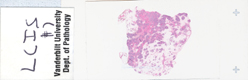
|
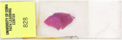
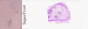
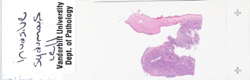
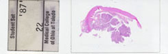
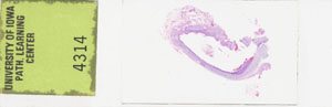
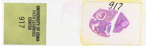
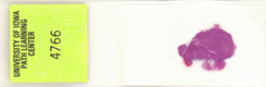
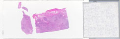
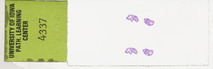
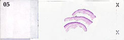
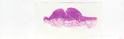
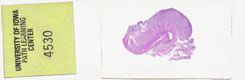
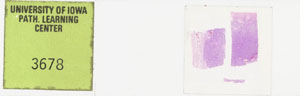
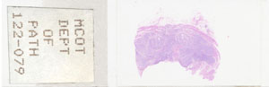
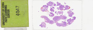
Abdomen--Desmoid tumor
Is this malignant? Demonstrate the entrapping of skeletal muscle fibers by
fibrous tissue.
Do you thing this could sometimes be mistakenly
diagnosed as malignant, even by experts?
Can it develop into a malignancy? If it did, would it
be a carcinoma or sarcoma? |
 |
|
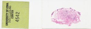
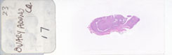
Adrenal--Adrenocortical carcinoma
Find some malignant appearing cells, using the usual nuclear
characteristics of malignancy, e.g., pleomorphism, hyperchromasia,
enlargement, etc. Describe the tumor cells in plain words rather than
those huge esoteric words I just mentioned. Are adrenocortical carcinomas
rare? Is it even rarer when they are "functional"? What does a functional
endocrine tumor mean? Is a benign tumor more likely to be functional? Why?
How do you know this might soon be ready to invade the kidney? |
 |
|
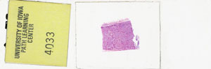
Adrenal--Cortical adenoma
Are these tumors common? What bright color are these grossly? What is the
difference between a functional and non-functional endocrine tumor? Could
they be either?
|
 |
|
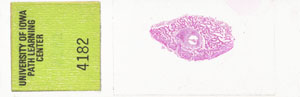
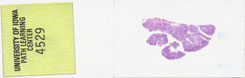
Adrenal--Neuroblastoma
Find a "rosette"? Is a neuroblastoma always used as a classical tumor to
show rosettes? Are neuroblastomas malignant? Is this one of the commonest
pediatric solid tumors? What is THE commonest?
|
 |
|
Adrenal--Nodular hyperplasia
Find a nodule. Could this be "functional"? Can you tell it is functional
just from the histology or would you need to measure hormone levels? What
is another way you could tell it is functional without doing any special
tests? (Hint: Look at the patient) Do the tumor cells look quite a bit
like "normal" adrenocortical cells? Is it a general principle, that if
2 cells look identical they may also behave identical?
|
 |
|
Adrenal--Pheochromocytoma
Why are these often called the most malignant looking benign tumors in
pathology? Does the cortex look normal? Could this produce a curable type
of hypertension? What is the diagnostic lab/physiologic test to prove this
tumor exists?
|
 |
|
Bladder--Urothelial carcinoma (transitional cell
carcinoma) in-situ
Find
an area of CIS. Is it associated with considerable underlying
inflammation? Does inflammation cause cancer?
|
 |
|
Bladder --Urothelial carcinoma Grade I
Why is this called Grade I?
Does it invade the
smooth muscle of the bladder wall? |
 |
|
Bladder --Urothelial carcinoma Grade III
Why is this classified as Grade III? Is a higher grade more likely to look
worse and behave worse?
Show bladder wall (i.e.,
smooth muscle) invasion. Can the words invasion and infiltration be used
interchangeably? |
 |
|
Blood--Chronic lymphocytic leukemia
What does a sustained peripheral smear lymphocyte count have to be in an
adult over 50 to warrant the diagnosis of CLL?
Do you need a bone marrow puncture to make this
diagnosis? Are these calls blasts or normal looking lymphocytes?
|
 |
|
Blood --Acute
leukemia
Find an absolutely indisputable blast cell, and describe. in
words, why it is a blast.
Does the correct identification of ANY TRUE blast
cell in a peripheral smear present suspicion for a leukemia? |
 |
|
Blood --Chronic myelogenous leukemia
Which of the three cell lines are classically elevated in this disease,
RBC, WBC, platelets, or all?
What percentage of
the myeloid series cells in this smear do you estimate to be blasts? Can
this turn into acute leukemia?
Which chromosome is classically abnormal in this
disease? Which large U.S. city is it named after? |
 |
|
Bone--Chondroma
Does a chondroma look like rather normal cartilage histologically? Is this
benign? If it wasn't benign, might it still look like normal cartilage?
Demonstrate lacunar "crowding". How many condrocytes
should normally live in a single lacuna?
|
 |
|
Bone--Fibrous dysplasia
Does the "fibrous" or the interspicular component of this tissue appear
"overgrown"? Do the spicules, conversely, look thinner than usual?
Show, i.e., dileanate a fibrous area between spicules?
Is this "dysplastic" in that it appears premalignant?
|
 |
|
Bone--Multiple myeloma
Find sheets of mostly normal looking plasma cells.
Should a normal marrow ever have "sheets" of plasma cells? What is the
normal acceptable percantage of marrow plasma cells? What is a monoclonal
gammopathy? Might this patient have one? |
 |
|
Bone--Osteoid osteoma
What is the "nidus" of an osteoid osteoma? Encircle it. Describe it in
words.
|
 |
|
Bone--Osteosarcoma
Like any sarcoma, what organ is this tumor most likely to metastasize to
first? Why, anatomically speaking?
|
 |
|
Bone-- Osteosarcoma
What is meant by the "bimodal" age distribution of osteogenic sarcoma?
Does tumor have more necrosis than the previous one? Find it.
|
 |
|
Bone marrow--adenocarcinoma,
metastatic
What is a signet ring cell? Could it be mistaken for a
plasma cell? Does it represent a poorly differentiated adenocarcinoma
cell? Attach a googled picture of a real signet ring, like the kind you
might wear on your finger.
|
 |
|
Bone Marrow--Chronic idiopathic myelofibrosis
Demonstrate the fibrosis of myelofibrosis? Should a normal adult marrow
have fibrosis? Is myelofibrosis associated with hematopoesis in other
organs which do not normally make marrow? Which 2 organs usually? What is
this phenomenon called? Is the spleen the most common site of
extramedullary hematopoeisis?
|
 |
|
Bone marrow --Follicular lymphoma
What percentage of the cells in the bone marrow are lymphocytes? What is
an acceptable normal percentage? Find some sheets of lymphocytes?
|
 |
|
Bone, vertebra--Adenocarcinoma, metastatic
Should true epithelial glands EVER be inside a bone marrow normally? If
you ever see any gland within a bone or marrow, what is the chances it is
metastatic adenocarcinoma rather than anything else?
|
 |
|
Brain--Astrocytoma
What percentage of CNS tumors are tumors of glial cells rather than tumors
of true neurons?
Can you find any true neurons in this tumor?
|
 |
|
Brain--Glioblastoma multiforme
What MAJOR pathologic finding has to be present in an astrocytoma before
you can call it a glioblastoma multiforme (GBM)? Find it. What are two
other common things which enable the diagnosis? Is this tumor also more
pleomorphic than the previous? Is it also more vascular?
What is the 1 year survival of an untreated GBM? |
 |
|
Brain-- Glioblastoma multiforme
What MAJOR pathologic finding has to be present in an astrocytoma before
you can call it a glioblastoma multiforme (GBM)? Find it. What are two
other common things which enable the diagnosis? Is this tumor also more
pleomorphic than the previous? Is it also more vascular?
What is the 1 year survival of an untreated GBM? Why might this
tumor have less CT density centrally? |
 |
|
Brain--Meningioma
Find a few of the numerous "psammoma" bodies. Are psammoma bodies fairly
diagnostic of meningiomas? Are most meningiomas "benign"?, i.e., do not
metastasize?
|
 |
|
Brain--Oligodendroglioma
Show some cells which look like normal oligodendral cells? It is
easy to figure out why these are called oligodendrogliomas?
|
 |
|
Brain, cerebellum --Medulloblastoma
What part of the cerebellum do these arise in, left lobe, right lobe, or
vermis? What age group do they arise in? Is a midline cerebellar tumor on
CT in an infant a medulloblastoma until proven otherwise?
|
 |
|
Brainstem--Glioma
Is a "glioma" a generic term for any brain tumor derived from any type of
glial cell? Name 5 types of glial cells?
|
 |
|
Breast--Colloid carcinoma
Find the "colloid"?
Is this also called a mucinous carcinoma? Is the
tumor a somewhat better prognosis than most other types of breast ductal
carcinomas?
|
 |
|
Breast--Ductal carcinoma in situ
Encircle an area of DCIS. Is "necrosis" a crucially important finding to
enable the diagnosis of DCIS (versus hyperplasia)?
|
 |
|
Breast--Fibroadenoma
Is this, by far, the most common well defined solid tumor in the breast of
young women?
Which is the more proliferated part
of the tumor, the glandular (epithelial) or stromal (connective tissue)
part of the tumor?
Are these more likely to be classified as stromal
rather epithelial tumors? Do they become cancer? Do they fibrose and
calcify with age? |
 |
|
Breast --Fibroadenoma
What percentage of this tumor is stromal rather than epithelial?
|
 |
|
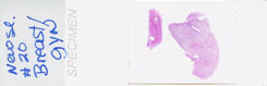
Breast--Gynecomastia
Is there differentiation into acini? Why not?
Name 2 common drugs which this is associated with.
|
 |
|
Breast--Intraductal papilloma
Does this papilloma invade into the duct wall or show necrosis? Why not?
Find some papillary projections.
|
 |
|
Breast--Lobular carcinoma
Find an "Indian file"? Is LCIS also present?
|
 |
|
Breast--Lobular carcinoma in situ
Does the finding of LCIS usually mandate a mastectomy? Find a lobule jam
packed with monotonous looking cells, i.e., they all look the same.
|
 |
|
Breast--Medullary carcinoma
What percentage of this tumor is benign lymphocytes? Find some malignant
glands. Is this type of ductal carcinoma also a somewhat
better-than-average prognosis?
|
 |
|
Breast--Paget disease
Find the "pagetoid" cells. In what percentage of these cases is there also
an underlying infiltrating malignancy?
|
 |
|
Breast --Ductal carcinoma
Are almost all breast carcinomas also called "ductal" carcinomas? Why?
Are almost all breast carcinomas also called "adeno" carcinomas? Why?
Is this ductal carcinoms "invasive" or "in-situ"
(i.e., DCIS)? |
 |
|
Breast --Fibroadenoma
Do fobroadenomas usually have the consistency of a superball? Is there any
calcification in this one?
Might there be
calcification if they took it out 30 years later? |
 |
|
Breast --Lobular carcinoma in situ
Find (encircle) a lobule with LCIS?
Describe it in words.
|
 |
|
Breast, soft tissue--Hemangioma
Can hemangiomas originate in any tissues or organs which have blood
vessels? Can they thrombose? Find a thrombosed area?
|
 |
|
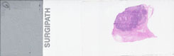
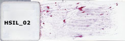
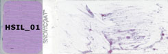
Cervix--High grade squamous intraepithelial
lesion (HSIL), Pap smear
Do "suspicious" cells often have enlargement, and increased N:C ratios and
hyperchromasia and pleomorphism? Find some.
|
 |
|
Cervix-- High grade squamous intraepithelial
lesion (HSIL), Pap smear
Does HSIL represent either severe dysplasia or malignancy usually? Find
some "suspicious" cells.
|
 |
|
Cervix -- High grade squamous intraepithelial
lesion (HSIL), Pap smear
Find some "suspicious" cells.
|
|
|
Cervix--HSIL
Find some "suspicious" cells.
|
 |
|
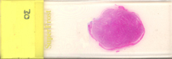
Cervix--Low grade squamous intraepithelial
lesion (LSIL), AutoCyte
Find some "mildly" suspicious cells, i.e., less enlargement, less
hyperchromasia, less pleomorphism, and lower N:C ratio than the HSIL.
|
 |
|
Cervix--Low grade squamous intraepithelial
lesion (LSIL), Pap smear
Find some "mildly" suspicious cells, i.e., less enlargement, less
hyperchromasia, less pleomorphism, and lower N:C ratio than the HSIL.
Has PAP screening cured, for all practical purposes, cervical cancer in
this country?
|
 |
|
Cervix --Low grade squamous intraepithelial
lesion (LSIL), Pap smear
Find some "mildly" suspicious cells.
|
|
|
Cervix--Squamous cell carcinoma
Find a nest of malignant invasive squamous cells. What is it invading?
|
 |
|
Cervix-- Squamous cell carcinoma
Can infiltrating SCC be confused with squamous metaplasia? What is the
difference?
|
 |
|
Cervix--Squamous metaplasia &
carcinoma-in-situ
Can tumor cells replace metaplastic cells within an
endocervical gland? Find the CIS. Is CIS also called CIN-III? Is CIN-III
also called severe dysplasia?
Find an area of "early" or "microscopic" invasion. Is this better
prognosis than an area of "advanced" or "extensive" invasion? |
 |
|
Cervix --Carcinoma-in-situ and early invasive
carcinoma
Find the area of
early invasion.
|
 |
|
Cervix -- High grade squamous intraepithelial
lesion (HSIL), Pap smear
Find some suspicious cells.
|
 |
|
Cervix --Low grade squamous intraepithelial
lesion (LSIL), AutoCyte
Find some mildly suspicious cells.
|
|
|
Cervix --Severe dysplasia, CIN III
Find some severely suspicious cells? Can the terms "severe dysplasia" and
"CIN-III" be pretty much used interchangeably?
|
 |
|
Cervix --Squamous cell carcinoma
Is this infiltrative?
Is this infiltrating?
Is this invasive?
Do all these questions mean the same? |
 |
|
Colon--Adenocarcinoma
Encircle the malignant mucosa on the left.
Encircle the benign mucosa on the right.
|
 |
|
Colon--Burkitt
lymphoma
Do 100% of all lymphomas have "effacement" of the normal nodal
architecture? Does the "normal nodal architecture" consist of follicles
under the cortical subcapsular sinus and medullary sinuses in the
medullary area? Please elaborate this crucially important concept? Is the
phrase "malignant lymphoma" redundant? Why? |
 |
|
Colon--Hyperplastic polyp
Do hyperplastic polyps turn into cancer?
Find
some "serrated" mucosal glands/ |
 |
|
Colon--Juvenile polyp
What is the definition of a hamartoma?
Is a juvenile polyp a classic example of a hamartoma?
Why?
|
 |
|
Colon--Tubular adenoma
Are the terms "adenomatous polyp" and "tubular adenoma", more or less,
synonymous?
Can these turn into cancer?
Find the "stalk". |
 |
|
Colon--Tubular adenoma (adenomatous polyp)
Are there any areas in which the glands are "back to back", i.e., no
connective tissue separating glands?
Why would it be hard to find this phenomenon here?
Is this tumor benign? Find the beautiful
fibrovascular "stalk". Does it have tumor in it? Why not? |
 |
|
Colon--Ulcerative colitis, carcinoma, atypia
Do many ulcerative colitis cases eventually develop into carcinoma?
What is the process called between normal mucosa and downright cancer
called? What is the definition of dysplasia?
Does dysplasia often take many years? Find a gland
with "atypia"? Is atypia synonymous with "dysplasia" from a histologic
point of view? |
 |
|
Colon--Villous adenoma
What percentage of villous adenomas eventually develop into cancer? What
percentage of tubular adenomas eventually develop into cancer?
Find "villous" (i.e. papillary or fingerlike"
structures.
Is the villous pattern of growth
more worrisome than the tubular pattern of growth? |
 |
|
Colon --Adenocarcinoma
Find some malignant glands invading the muscularis layer.
|
 |
|
Colon --Adenomatous polyp (tubular adenoma) with
a focus of carcinoma
Encircle the part of the polyp that has turned into cancer. Describe, in
words, how it differs from the benign part.
|
 |
|
Colon --Tubular adenoma (adenomatous polyp)
Is the fibrous stalk nice and clear of tumor glands. If the stalk DID have
glands in it, would it help you to decide between malignancy and
benignancy?
|
 |
|
Diaphragm--Metastatic liposarcoma
What organ do all sarcomas metastasize to first? Why?
Should a diaphragm be 100% skeletal muscle and/or
dense fibrous connective tissue?
Is this one
normal? Why/Why not? |
 |
|
Endometrium--Endometrial polyp
What causes benign endometrial polyps? Is the underlying myometrium
invaded?
Draw a nice line delineating myometrium
from endometrium. |
 |
|
Esophagus--Adenocarcinoma
Is "adenocarcinoma" the second most common type of esophageal carcinoma?
What is #1?
Find some malignant glands invading smooth muscle. |
 |
|
Esophagus--Squamous cell carcinoma
Find some invasive SCC.
Find some in-situ SCC.
|
 |
|
Esophagus, liver--Squamous cell carcinoma with
metastases
Find metastatic
nests within lung spaces? Are these spaces lymphatics, veins, arteries, or
alveoli?
|
 |
|
Extrahepatic bile ducts, common bile
duct--Adenocarcinoma, moderately differentiated
Name 4 different extrahepatic bile ducts.
Find the cancer (i.e., adenocarcinoma).
Does it look "papillary" as well? |
 |
|
Eye --Retinoblastoma
Do the central portions of these dark tumor cell nests contain blood
vessels?
Do the parts NOT fed by blood vessels look
"necrotic"? Why?
What happens to a tumor that is growing so fast that
it outgrows its own blood supply? |
 |
|
Heart, pericardium--Metastatic breast
carcinoma
Find a tumor
nest in pericardium.
Should pericardium normally have true epithelial
glands?
What primary tumor most often
metastasizes to pericardium? Why? |
 |
|
Kidney--Renal cell carcinoma
Delineate the tumor. Delineate the normal kidney.
Why is renal cell carcinoma often called clear cell
carcinoma? Do the tumor nuclei look rather benign appearing?
Is renal cell carcinoma more likely to metastasize to lymphatics first or
veins? |
 |
|
Kidney-- Renal cell carcinoma
What percentage of this tumor is composed of blood vessels? Would you
expect this to light up like a light bulb on angiography?
|
 |
|
Kidney--Renal cell carcinoma (hypernephroma)
Is hypernephroma genarally synonymous with renal cell carcinoma?
Do the typical tumor cells have a central small well
defined nucleus and foamy (i.e., "clear") cytoplasm? Why are these also
called clear cell carcinomas? Verify clear cells by showing some. Does the
foamy clear cytoplasm contain glycogen?
Can you
find any tumor cells in veins? |
 |
|
Kidney--Transitional cell carcinoma
Can you find any tumor cells in veins here? Why not? Which part of the
kidney gives rise to TCC, cortex, medulla, or pelvis?
Does the tumor look papillary? Show some papilla.
|
 |
|
Kidney--Wilms tumor
Is this the most common pediatric solid malignant tumor? What is it's
survival in 2006? What was it in 1956?
Because
this is a kidney tumor, do you thing it might be trying to form some
primitive glomeruloid structures? Find some. |
 |
|
Liver--Acute myeloid leukemia
Find blasts in the liver. Are they portal or centrilobular? Could these be
ANY type of hematopoetic blast cell?
|
 |
|
Liver--Cirrhosis, hepatocellular
carcinoma
Do hepatomas often, or usually, develop in a background of
cirrhosis?
Can a "nodule" of cirrhosis often be
indistinguishable from a tumor nodule of hepatoma, even by an expert?
Find the cirrhosis area. Find the hepatoma. Describe
the difference in plain English. |
 |
|
Liver--Colangiocarcinoma, moderately well
differentated
What are
the only glandular structures normally found in a liver?
So a primary adenocarcinoma of the liver would have to be derived from the
_______? Find some cholagiocarcinoma glands.
What percentage of adenocarcinomas occupying the
liver are primary to the liver rather than metastatic? |
 |
|
Liver--Hepatocellular carcinoma in cirrhosis of
the liver
Find the tumor
area on top.
Find the cirrhosis area on the bottom.
In your own words, how would you describe the difference? |
 |
|
Liver--Metastatic adenocarcinoma of stomach
Can we tell if this metastatic tumor originated in the stomach just by the
microscopic appearance?
Find some metastatic
nests of tumor cells? |
 |
|
Lung--Adenocarcinoma
Are adenocarcinomas of
the lung generally located more peripheral than the squamous cell
carcinomas? Why?
Find some malignant glands. |
 |
|
Lung--Bronchial carcinoid
Are carcinoid cells neuroendocrine cells? Where are
neuroendocrine cells derived from embryologically?
Are "oat" cells also neuroendocrine cells? What is an
"oat" cell? Is oat cell carcinoma the worst type of lung cancer?
What is another name for oat cell carcinoma?
What % of this slide is tumor? Delineate tumor from normal bronchial wall. |
 |
|
Lung--Bronchiolo-alveolar carcinoma
Encircle the tumor. Is this type of primary adenocarcinoma of the lung
much slower growing than other types?
|
 |
|
Lung--Hamartoma
Define hamartoma in general. Are most primary lung hamartomas chiefly
cartilage?
|
 |
|
Lung--Metastatic adenocarcinoma
Encircle the met.
Are most adenocarcinoma foci
in the lung guessing games as to where they came from, without knowing a
patient's history? |
 |
|
Lung--Metastatic leiomyosarcoma
What percentage of this slide is tumor? How might this
present clinically?
If this patient was female, where would be the most
likely source? How many uterine leiomyomas turn into sarcomas?
What is the usual first symptom in a patient with a
metastatic sarcoma? |
 |
|
Lung--Small cell carcinoma
What is the old name for this cancer?
Are the
nuclei, small, fragile, and appear often necrotic with minimal cytoplasm?
Show some. Use your own words to describe it as well. Is this the only
type of primary lung cancer which has the best response to chemotherapy,
even though, without treatment, it has the worst prognosis? How important
is it then to distinguish "small cell" from "NON small cell" carcinoma on
a small needle biopsy? |
 |
|
Lung (view 1), bone (view 2),liver (view 3) --Small cell carcinoma with metastases
Find some small cell carcinoma nests.
|
 |
|
Lung --Squamous cell
carcinoma
In what part of the lung does SqCC start. Could it be
preceded, usually, for many years by squamous dysplasia in a bronchus?
Show some SqCC invading bronchus? |
 |
|
Lung, bronchus--Carcinoma in situ
If these malignant cells actually invaded through bronchial wall, would
you still call it CIS?
Find some CIS?
|
 |
|
Lung, esophagus--Squamous cell carcinoma with
metastasis
Which side of
the bronchial cartilage is the tumor, above, below, or both?
|
 |
|
Lung, lymph node --Bronchioloalveolar carcinoma
with metastasis to lymph node
What % of the lymph node on the left has tumor?
What % of the lung on the right has tumor?
Which part of a normal lymph node has the earliest
focus of metastasis? Why? |
 |
|
Lung, pleura--Mesothelioma
If this tumor was not grossly confined to the pleura, could it be easily
mistaken for primary non-small cell lung cancer (i.e., adenocarcinoma,
squamous carcinoma, or large cell carcinoma)?
Do
you think there are a whole bunch of cellular antigens we can stain for to
help us determine whether this is a mesothelioma or NON-small cell
carcinoma? |
 |
|
Lung, pleura-- Mesothelioma
Which pleura is giving rise to this mesothelioma, visceral or parietal?
Does it also have kind of a "papillary" appearance?
|
 |
|
Lung, subpleural--Metastatic small cell
carcinoma
Encircle the
metastatic area? Describe what "small cells" look like in plain English.
|
 |
|
Lymph node--Anaplastic large cell
lymphoma
Is 100% of this lymph node's "normal architecture"
effaced and replaced by lymphoid cells?
Is this the underlying process in all lymphomas, no
matter what lymphocyte antigens (i.e., "markers") you have fun staining
for?
Describe what the "normal architecture" is of a lymph
node. |
 |
|
Lymph node--Chronic lymphocytic leukemia
- small
lymphocytic lymphoma
Is 100% of this lymph node's "normal architecture"
effaced and replaced by lymphoid cells?
Is this the underlying process in all lymphomas, no
matter what lymphocyte antigens (i.e., "markers") you have fun staining
for?
Describe what the "normal architecture" is of a lymph
node. |
 |
|
Lymph node--Langerhans cell histiocytosis
(Hand-Schuller-Christian disease)
Is 100% of this lymph node's "normal architecture"
effaced and replaced by lymphoid cells?
Is this the underlying process in all lymphomas, no
matter what lymphocyte antigens (i.e., "markers") you have fun staining
for?
Describe what the "normal architecture" is of a lymph
node. |
 |
|
Lymph node--Lymphoblastic
lymphoma - lymphoblastic leukemia
Is 100% of this lymph node's "normal architecture"
effaced and replaced by lymphoid cells?
Is this the underlying process in all lymphomas, no
matter what lymphocyte antigens (i.e., "markers") you have fun staining
for?
Describe what the "normal architecture" is of a lymph
node. |
 |
|
Lymph node--Lymphoplasmacytic
lymphoma
Is 100% of this lymph node's "normal architecture"
effaced and replaced by lymphoid cells?
Is this the underlying process in all lymphomas, no
matter what lymphocyte antigens (i.e., "markers") you have fun staining
for?
Describe what the "normal architecture" is of a lymph
node. |
 |
|
Lymph node--Mantle cell
lymphoma
Is 100% of this lymph node's "normal architecture"
effaced and replaced by lymphoid cells?
Is this the underlying process in all lymphomas, no
matter what lymphocyte antigens (i.e., "markers") you have fun staining
for?
Describe what the "normal architecture" is of a lymph
node. |
 |
|
Lymph node--Metastatic breast carcinoma
Is "partial" effacement of a lymph node common with metastatic carcinoma?
|
 |
|
Lymph node--Nodal marginal zone B-cell
lymphoma (with plasma cell differentiation)
Is 100% of this lymph node's "normal architecture"
effaced and replaced by lymphoid cells?
Is this the underlying process in all lymphomas, no
matter what lymphocyte antigens (i.e., "markers") you have fun staining
for?
Describe what the "normal architecture" is of a lymph
node. |
 |
|
Lymph node--Nodular sclerosis Hodgkin
lymphoma
What are the two kinds of malignant lymphomas? Is there such a
thing as a benign lymphoma?
Does the finding of a Reed Sternberg cell enable us
to differentiate a Hodgkins lymphoma from the other type of lymphoma,
which is called a non-Hodgkins lymphoma? If so, you better learn how to
identify them. Identify some. What percentage of all lymphomas are
Hodgkins lymphomas? |
 |
|
Lymph node--Peripheral T-cell lymphoma,
unspecified (mixed medium-sized and large
cell)
Do the infiltrates from CLL look like rather normal looking
lymphocytes?
Can anybody tell the difference between a lymph node
replace by CLL cells and a lymphocytic lymphoma? |
 |
|
Lymph node --Adenocarcinoma, metastatic to lymph
node
What % of this lymph
node is replaced by tumor? Is there any normal lymph node left? If so,
where and how much?
|
 |
|
Lymph node --Diffuse large B-cell
lymphoma
Is 100% of this lymph node's "normal architecture"
effaced and replaced by lymphoid cells?
Is this the underlying process in all lymphomas, no
matter what lymphocyte antigens (i.e., "markers") you have fun staining
for?
Describe what the "normal architecture" is of a lymph
node.
How many different, stupid, classifications of
lymphomas have we allowed ourselves to put up with over the years? |
 |
|
Lymph node --Follicular lymphoma, grade
2
Is 100% of this lymph node's "normal architecture"
effaced and replaced by lymphoid cells?
Is this the underlying process in all lymphomas, no
matter what lymphocyte antigens (i.e., "markers") you have fun staining
for?
Describe what the "normal architecture" is of a lymph
node. |
 |
|
Lymph node --Metastatic undifferentiated large
cell carcinoma
Find an
area in which you can show both carcinoma cells (in sheets), as well as
lymphoid cells?
What is the main basic
difference between a carcinoma cell and a lymphoma cell, embryologically
and morphologically? |
 |
|
Lymph node, bone marrow, liver, kidney--Chronic
lymphocytic leukemia - small lymphocytic lymphoma
Do all the infiltrates of CLL look like normal looking lymphocytes
usually?
Find some. |
 |
|
Mouth--Pyogenic granuloma
In all honesty, can a pyogenic granuloma also be called "granulation
tissue"? What is the single most dominant feature of granulation tissue
histologically? Identify this.
|
 |
|
Neck--Lymphangioma (cystic hygroma)
The lack of what kinds of cells in the lumen of the endothelially lined
spaces makes us suspect lymphangioma rather than henamgioma?
|
 |
|
Neck --Branchial cyst (lymphoepithelial
cyst)
Description Cysts are benign tumors lined by
benign epithelium. They are filled with epithelial cell secretions and/or
sloughed cellular debris. Although they are "tumors" they are usually not
neoplasms in the strict sense; instead they usually arise from epithelial
inclusions that do not have access to the surface. (For info
only, but describe the cyst wall) |
 |
|
Nose--Extramedullary plasmacytoma
What does "extramedullary hematopoesis" mean conceptually? Which is the #1
organ which might have this? Which is the #2 organ?
If you saw extramedullary hematopoesis in a spleen or liver, might you
suspect something might be going on on the marrow which is making it
difficult for hematopoesis to go on there, in it normal place? Where
are blood cells made in the fetus? |
 |
|
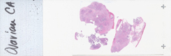
Ovary-- Adenocarcinoma
Are most ovarian cancers adenocarcinomas? Do these cells look like they
are more likely to be making clear fluid (i.e., serous), rather than
mucinous fluid? Find some serous papillary structures.
|
 |
|
Ovary--Adenocarcinoma
Every time you see an ovarian cancer, ask your self three questions: Is it
serous or mucinous, Is it solid or cystic, Is it papillary or not?
Show, and tell, which one this is. Do the three types
of ovarian cancers classifications depend on whether they originate from
the surface "Mullerian" epithelium, the germ cells, or the stroma? Which
type is by far the most common?
|
 |
|
Ovary--Benign teratoma
Is this the most common type of true ovarian neoplasm in young women? What
is another name for it?
Are the terms "dermoid cyst" and "benign cystic
teratoma" synonymous?
|
 |
|
Ovary--Borderline ovarian tumor
Does "borderline" mean the true biologic behavior cannot be 100% reliably
predicted from its microscopic appearance, or the pathologist can't make
up his mind? Does it have some papillae? Find some.
|
 |
|
Ovary--Brenner tumor
Is this an epithelial or a germ cell derived tumor?
Find some Brenner tumor cell nests.
|
 |
|
Ovary--Dermoid cyst
What is another name for this?
What two things
are almost always found inside the cyst wall? Find both. What are other
common things?
Why are they cysts greasy when one opens them up? |
 |
|
Ovary--Dysgerminoma
If this was a testicle rather than an ovary, what would you call it?
Find the malignant germ cells. Find some lymphocytes?
Are various mixtures of malignant germ cells and
lymphocytes also the exact same components of seminomas of the testis? |
 |
|
Ovary--Endometriosis
Identify the endometrial tissue in the ovary? Is this one of the most
common sites for endometriosis? What are some others?
Define endometriosis. Explain the embryologic rest cell theory vs. the
reverse menstruation theory/? Has anybody ever found out for sure
what causes this? |
 |
|
Ovary--Granulosa cell tumor
What might the effect of a granulosa cell tumor of the ovary be on the
endometrium? Why?
What do granulosa cells normally make?
|
 |
|
Ovary--Serous
cystadenocarcinoma
Why is this called "serous"? papillary"
cystic? carcinoma? adeno?
Find some papillary structures? Find cysts. |
 |
|
Ovary--Serous
cystadenoma
What is the diagnostic and predictive importance of the
word "borderline" on a pathology report? Why does this cause anxiety for
the patient and the surgeon? |
 |
|
Ovary--Theca cell tumor
What do theca cells normally produce? What might this tumor produce if it
is "functional"?
Encircle the part of the tumor
which might produce estrogen. Would a benign thecal tumor be more likely
to produce estrogen than, say, a theoretical malignant one? |
 |
|
Pancreas--Adenocarcinoma
Are most of the cancers of the pancreas adenocarcinomas? Why? Could a
squamous cell carcinoma be possible? How?
Does extreme fibrous reaction to this tumor, i.e., "desmoplasia",
occur routinely with pancreatic adenocarcinomas and breast carcinomas?
Which organ receives venous blood drainage from the
pancreas? Which organ gets the first metastasis from
pancreatic cancers? |
 |
|
Pancreas --Adenocarcinoma
Find normal pancreas.
Find a desmoplastic tumor area.
Find malignant glands within the desmplastic area.
Define desmoplasia.
What does the Greek word
desmos mean? |
 |
|
Pancreas --Islet cell tumor (insulinoma)
Find a normal islet cell.
Find an islet cell tumor cell.
Do they look virtually identical? Do you think they
might behave identical also, functionally? What do normal
islet calls make?
If these normal looking islet cells from the tumor
spread to the liver, would you have a hard time deciding whether it was
benign or malignant? |
 |
|
Parathyroid--Oxyphil adenoma
What is the normal weight of a single parathyroid gland?
Which part of the tissue section has almost 100% oxyphils, NW, NE, SE, or
SW quadrant? Are tumors generally "clonal"? What does that mean? |
 |
|
Penis--Carcinoma in situ
Find an area of squamous cell CIS.
|
 |
|
Penis--Erythroplasia of Queyrat
Is this also CIS? Find it.
|
 |
|
Penis--Squamous cell carcinoma
Is this CIS or infiltrating carcinoma or both or neither?
Find the deepest level of infiltration. |
 |
|
Pituitary--Acidophilic adenoma
What hormones do pituitary acidophils normally produce? What hormone
might this tumor produce? Name 2 ways you can determine whether a
pituitary tumor is functional or not---one lab way, and one clinical way.
What clinical syndrome might a functioning acidophilic adenoma produce?
What do basophils produce? What do chromophobes produce? |
 |
|
Pre-sacral mass --Diffuse large B-cell lymphoma
(needle biopsy)
Do you have enough experience by now to suspect this is
a lymphoma rather than a carcinoma or a sarcoma? (Honest answers
only, please) |
 |
|
Prostate--Adenocarcinoma
In what quadrant of this section is the main bulk of the adenocarcinoma,
NW, NE, SE, SW?
Are the overwhelming majority of
prostate cancers adenocarcinomas? Why? |
 |
|
Prostate--Adenocarcinoma (Gleason grade 1)
Do you agree with this Gleason grade? Why?
|
 |
|
Prostate--Adenocarcinoma (Gleason grade 4)
Do you agree with this Gleason grade? Why?
|
 |
|
Prostate--Adenocarcinoma (Gleason grade 5)
Do you agree with this Gleason grade? Why?
|
 |
|
Prostate--Intraepithelial neoplasm (PIN)
Find the PIN.
Can PIN be thought of as the spectrum of precancerous
changes leading up to downright adenocarcinoma?
What percentage of men over 80 have prostate cancer or PIN? |
 |
|
Prostate --Adenocarcinoma (Gleason grade 2)
Do you agree with this Gleason grade? Why?
|
 |
|
Prostate --Adenocarcinoma (Gleason grade 3)
Do you agree with this Gleason grade? Why?
|
 |
|
Prostate --Nodular
hyperplasia
Why is this called "nodular"? Find a nodule? Do the cells
look malignant?
What is the MAIN feature differentiating hyperplastic
glands in the prostate, from cancer, pleomorphism, nucleoli, or necrosis? |
 |
|
Salivary gland--Adenoid cystic carcinoma
Show a malignant area. Show a necrotic one. Are these tumor glands slow
growing and do they invade perineural spaces frequently?
Is this the classic example of a widespread metastatic malignancy which a
patient can live with for many many years because of its extreme slow
growth rate? |
 |
|
Salivary gland--Extranodal marginal zone B-cell
lymphoma of mucosa-associated lymphoid tissue (MALT lymphoma)
What does MALT stand for? Why?
Can MALT tissue give rise to lymphomas? What is a
maltoma? Is a maltoma a lymphoma of extranodal lymphoid tissue?
Find an area of lymphoid cells in which there is not
much further organization that simply lymphoid cells? |
 |
|
Salivary gland--Papillary cystadenoma
lymphomatosum (Warthin tumor)
Is this malignant? Find the cyst part. Find the lymph part. Find the
papillary part? Why is it more truly descriptive of this tumor to call it
a papillary cystadenoma lymphomatosum rather than a Warthin tumor?
|
 |
|
Salivary gland--Pleomorphic adenoma (mixed
tumor)
Is this the most
common, by far, salivary gland neoplasm? Is it benign? Delineate the edges
of it. From the sharp delineation of the tumor from its neighboring
structures, would you guess it's benign? What is the Latin word for
"crab"? Do crabs have claws which invade neighboring structures? Why is
cancer called cancer?
|
 |
|
Skeletal muscle, face--Alveolar
rhabdomyosarcoma
What
normal cells are rhabdomyosarcoma cells derived from? Are there rare? What
organ usually receives the first met?
|
 |
|
Skin-- Actinic
keratosis
Is this regarded as the premalignant precursor to squamous
cell carcinoma of the skin, generally? Is it caused by sun exposure?
Find the hyperkeratosis? What is hyperkeratosis? Find
the atypia. What is atypia? Are the words "atypia" and "dysplasia" sort of
the same? |
 |
|
Skin--Actinic keratosis
Is this regarded as the premalignant precursor to
squamous cell carcinoma of the skin, generally? Is it caused by sun
exposure?
Find the hyperkeratosis? What is hyperkeratosis? Find
the atypia. What is atypia?
|
 |
|
Skin--Dermatofibroma
What is the cell of
origin of a dermatofibroma? Is this sometimes called a "histiocytoma"? Is
it sometimes called a sclerosing hemangioma?
Are the tumor cells very hard to differentiate from
surrounding fibroblasts? Try to anyway. |
 |
|
Skin--Epidermal inclusion
cyst
What is the most common subcutaneous "bump" on a person's skin?
What are they filled with? Can they rupture? Find a giant cell trying to
swallow some ruptured keratin. |
 |
|
Skin--Fibroepithelial polyp (skin tag)
Are these common? Find one on your own body. Where would you look first?
Do they turn into cancer? What causes them?
Find the "fibro" part. Find the "epithelial"
part.
|
 |
|
Skin--Glomus tumor
Does a glomus tumor look like it might be derived from blood vessels, just
from looking at this image?
|
 |
|
Skin--Granular cell tumor
Find some "granular" cells. What are these granules made of? What is a
better stain than H&E to see these granules? What is a "supravital" stain?
Is methylene blue a supravital stain?
|
 |
|
Skin--Kaposi sarcoma
IS KS common in non-AIDS patients?
Is it much more common in homosexual HIV patients
rather than IVD user AIDS patients?
Is it derived
from blood vessels? Is it malignant? Where do sarcomas metastasize to? |
 |
|
Skin--Keratoacanthoma
What malignancy is KA most commonly mistaken for, even by experts, often?
Why?
Does the presence of an epithelial "collar"
or "collarette" at he\\the periphery help us to differentiate the two?
Find the collarette. |
 |
|
Skin--Melanoma in situ
Do melanomas start off "in-situ"?
Find an area of increased melanocytes. Does the
presence of melanocytes in the upper epidermis present a worrisome
finding?
|
 |
|
Skin--Mycosis
fungoides
Is MF a type of cutaneous T-Cell lymphoma? Are there stains
to prove these are T-Cells? Find some sheets of tumor cells. Can you tell
any lymphocyte is a T-Cell or a B-Cell just from routine stains? |
 |
|
Skin--Neurofibroma
Is it common to find neurofibromas sporadically, with no association with
hereditary neurofibromatosis syndromes?
Describe
the tumor cells in plain English?
Do they look malignant? Are neurofibromas malignant?
Does the overlying epidermis look pretty normal? |
 |
|
Skin--Palisaded Encapsulated Neuroma (solitary
neuroma)
What is the
definition, in pathology or anywhere, of "palisading"? Find some here.
If you see a golden brown pigment in the cells any primary skin tumor,
what are the chances it is bile? Hemosiderin? Melanin? |
 |
|
Skin--Pigmented Spindle Cell Nevus
Do these pigmented cells have a "spindly", or fusiform, appearance?
|
 |
|
Skin--Schwannoma
Do these tumor cells look spindly? Can you find "palisading"? Does
it look malignant by any criteria?
Does the
number of sebaceous gland here look increased relative to all the other
skin specimens you have looked at histologically? |
 |
|
Skin--Sebaceous hyperplasia
Which part of the body is subject to sebaceous hyperplasia? Which part of
the body has the most sebaceous glands? What is seborrhea, clinically?
|
 |
|
Skin--Seborrheic keratosis
Find a trapped area of "whoorled" keratin? What is another name for this?
Are these fairly diagnostic of SK?
|
 |
|
Skin-- Seborrheic keratosis
What are "horn cysts"? Find some. Do these turn into cancer? Is SK related
to sun exposure, as AK (actinic keratosis) is?
|
 |
|
Skin--Squamous cell carcinoma
How do you know this is malignant?
How do you know this is derived from squamous
epithelium?
|
 |
|
Skin-- Squamous cell carcinoma
Do SqCCs of the skin often spread to distant organs?
Have you ever personally heard of any person dying from a squamous cell
carcinoma of the skin? |
 |
|
Skin or oral mucous membrane--Pyogenic
granuloma
Describe granulation tissue in words.
Describe a pyogenic granuloma in words.
What is the difference? |
 |
|
Skin --Malignant melanoma
Why is this the most feared Skin cancer? Do they often metastasize early
and widely? Are they on the rise, significantly?
What characteristic of a melanoma is the prognosis DIRECTLY related to:
vertical thickness, diameter, necrosis, or atypia?
Find the deepest level of dermal invasion. Is this
the most reliable histological criteria for melanoma prognosis? |
 |
|
Skin, lip--Basal cell carcinoma
Is BCC the overwhelmingly most common type of skin cancer? Do they
metastasize widely? Find the palisading of the basal cells?
Have you ever personally heard of any person dying from a basal cell
carcinoma of the skin? |
 |
|
Skin, nose--Rhinophyma
What famous comedian of the distant past could be the rhinophyma poster
boy?
Describe it microscopically.
|
 |
|
Small intestine--Carcinoid tumor
Find the carcinoid cells. Are the neuroendocrine cells? In the GI tract do
they look a lot like adenocarcinomas? Show them invading the muscularis.
If they invade the muscularis, do you think this one could metastasize
too? Would you call this one malignant for that reason? If the cells look
100% benign but you found them metastasizing to the liver, would you
modify your diagnosis from benign to malignant?
|
 |
|
Small intestine, duodenum--Peutz-Jeghers
polyp
What is a
Peutz-Jeghers polyp? What type of tissue does this polyp have that
differentiates it from regular adenomatous polyps? Find some smooth
muscle. Is this benign? Does it look benign?
|
 |
|
Soft tissue--Fibrosarcoma
What common connective tissue cells are fibrosarcomas derived from?
What is, unfortunately, the usual site of
presentation of any sarcoma?
|
 |
|
Soft tissue--Lipohemangioma
Why is this called both a "lipo" and a "hemangio"? Find the "lipo" part.
Find the "hemangio" part. Is this benign?
|
 |
|
Soft tissue (retroperitoneum) --Liposarcoma
What part of the body do most liposarcomas arise in? Does this look like
histologically normal adipose tissue? If it did, would you consider
changing your diagnosis to benign lipoma? Is the term benign lipoma
redundant?
|
 |
|
Soft tissue --Lipoma
Does a lipoma look like 100% normal adipose tissue microscopically? Does
it often have a thin fibrous capsule however?
If this was just a piece of normal fat would you
believe it?
|
 |
|
Soft tissue, dorsal wrist--Ganglion
What part of a tendon do ganglions arise from? Are they usually cystic? Do
they usually contain this mucoid material? Can this also be called a
benign "synovial" cyst?
|
 |
|
Stomach--Adenocarcinoma
Which of the 4 classical GI wall layers is involved by tumor? All?
Are there any signet ring cells? Find some if you can see any.
|
 |
|
Stomach--Adenocarcinoma (linitis plastica)
Where does the term linitis plastica come from? Is this synonymous with a
poorly differentiated adenocarcinoma involving the full thickness of a
stomach so that it looks like a leather bottle? Could there be signet ring
cells here? Find some if you can.
|
 |
|
Tendon sheath--Giant cell tumor
Are these benign? Find the giant cells which are very numerous. Is tendon
sheath a synovial derived structure also? What are some others?
|
 |
|
Testis--Embryonal
carcinoma
Is embryonal carcinoma the most often testicular tumor
erroneously referred to as "adenocarcinoma"? Why? What tumors metastasize
to the testis (Correct answer: NONE). Where do testicular cancers
metastasize to, inguinal or periaortic (retroperitoneal) lymph nodes? Why?
What is the old erroneous name for this cancer?
Circle some areas that look like adenocarcinoma. |
 |
|
Testis-- Embryonal carcinoma
What percentage of the tumor looks like glands? Is embryonal carcinoma a
"germ cell" tumor of the testis? Name 3 others? Are they all malignant?
What % is necrotic?
|
 |
|
Testis--Endodermal sinus tumor (yolk sac tumor)
Can you find a
Schiller-Duval body which is diagnostic of this tumor? If not, google one.
|
 |
|
Testis--Seminoma
If this was an ovary what would your diagnosis be?
These tumors are composed of 2 types of cells, aren't
they?
Find the malignant germ cells. Find the lymphocytes.
Which cell is more common in this case? |
 |
|
Testis-- Seminoma
Find the malignant germ cells. Find the lymphocytes. Which one is more
abundant in this one? Can they be 50-50?
|
 |
|
Testis--Sertoli cell tumor
What do sertoli cells normally produce? What might a functional sertoli
tumor also produce?
|
 |
|
Testis --Benign teratoma
Are most teratomas of the testis malignant?
Are
most teratomas of the ovary benign?
Interesting, isn't it? |
 |
|
Thymus--Thymoma
What common neuromuscular junctional autoimmune disease of women, mostly,
is this tumor associated with?
Are thymectomies common in myasthenia gravis?
From you experience does it look pretty much like a lymphoma? If this
tumor was taken out of an inguinal area what would most pathologists
diagnosis it as? |
 |
|
Thyroid--Follicular carcinoma
Find some malignant follicles. What is the usual route of early metastasis
for a follicular carcinoma? papillary?
What four malignancies often metastasize to
bone early?
Have you ever heard of somebody dying from thyroid
cancer? Have you ever personally heard of a male getting thyroid cancer? |
 |
|
Thyroid--Hashimoto thyroiditis, papillary
carcinoma
Find the
papillary structures? Are ALL papillary structures in the thyroid
regarded, pretty much, as being malignant?
Google and past a picture of Orphan Annie? Why did I ask you to do that?
Find an "Orphan Annie" cell of thyroid cancer, either from here or google? |
 |
|
Thyroid--Nodular goiter
What is the definition of a goiter? What is the normal weight of an adult
thyroid gland, in grams? What is the normal range of a 24 hour uptake of
radioactive iodine, expressed as a percentage of the entire dose? Amazing
coincidence, isn't it?
Find a nodule by
encircling it. Is the capsule "intact" and not disrupted in any way? Why
is this important? |
 |
|
Thyroid --Follicular adenoma (microfollicular
variant)
Can may thing
which look like normal follicular "adenomas" metastasize to bone and
liver? If it did would you change your diagnosis from benign to malignant?
Find a "micro"-follicle.
|
 |
|
Thyroid --Papillary carcinoma
Find some papillae. What percentage of all papillary structures in the
thyroid are eventually called "carcinoma"? What kind of cell is produced
in a thyroid when a cytoplasm invaginates into a nucleus? What famous
comic book orphan is this named after?
|
 |
|
Tongue--Squamous cell carcinoma
Is this infiltrating? Find some tumor cells choking skeletal muscle
fibers.
Does it look squamous? Why?
What is a squamous "pearl"? Find one. |
 |
|
Tongue-- Squamous cell carcinoma
Is this infiltrating?
Does it look squamous? Why?
What is a squamous "pearl"? Find one if you see one. |
 |
|
Ureter--Transitional cell
carcinoma
Are virtually all primary malignancies of the male urogenital
tract classified as TCC? What is another name for TCC? Does this one
invade the smooth muscle of the ureter? Show it. |
 |
|
Urinary bladder--Transitional cell carcinoma
Does this one invade the smooth muscle of the bladder? Show it.
|
 |
|
Uterus--Adenomyosis
What is the definition of adenomyosis? Why is it also called endometriosis
interna? Do you think it would be better to see BOTH endometrial gland and
stroma in the myometrium to diagnose this condition? Find some.
|
 |
|
Uterus--Choriocarcinoma
Do you see anything here which reminds you of chorionic villi?
Syntrophoblast? Cytotrophoblast? If so, show it.
What hormone does this tumor usually secrete?
|
 |
|
Uterus--Endometrial adenocarcinoma
Are virtually all of the primary carcinomas of the endometrium
adenocarcinomas? Why? Show the depth of invasion.
|
 |
|
Uterus--Leiomyoma
Is this the most common tumor of women? Where do most of them originate
in? Microscopically, does it look pretty much like normal smooth
muscle histologically?
|
 |
|
Uterus--Leiomyosarcoma
What percentage of leiomyomas turn into leiomyosarcomas? What SINGLE
cytologic feature distinguishes leiomyomas from leiomyomasarcomas,
mitoses, atypia, necrosis, or fibrosis? Take 10 HPFs (High Power Fields).
Count the total number of mitotic cells in those 10 fields.. What is that
number? _______ What should the number be in a benign leiomyoma?
|
 |
|
Uterus--View 1 Endometrial adenocarcinoma, View
2 Fibromyoma (leiomyoma)
Find the adenocarcinoma. Find the non-malignant endometrium.
Find the myometrium? Is it invaded?
|
 |
|
Uterus --Leiomyoma
Delineate it.
|
 |
|
Uterus --Leiomyosarcoma
What SINGLE cytologic feature distinguishes leiomyomas from
leiomyomasarcomas, mitoses, atypia, necrosis, or fibrosis? Take 10 HPFs
(High Power Fields). Count the total number of mitotic cells in those 10
fields.. What is that number? _______ What should the number be in a
benign leiomyoma? |
 |
|
Uterus, corpus, placenta--Hydatidiform mole
Do these look like normal chorionic villi, or do they seem to be bloated
with edema so the look like grapes?
What is the
Greek ford for water? Latin? |
 |
|
Uterus, endometrium--Adenocarcinoma
Could this be a D&C specimen? What does D&C stand for? Are D&C's performed
often to rule out cancer as a cause of abnormal uterine bleeding? Can we
rule it out here? Can we rule it in?
|
 |
|
Vagina--Clear cell carcinoma
Why is this pattern of adenocarcinoma often called "clear" cell carcinoma?
Find some clear foamy tumor cells.
|
 |
|
Vulva--Severe dysplasia
Would call this VIN-I, VIN-II, or VIN-III? Why? Encircle an area?
|
 |
|
Vulva --Condyloma accuminata (viral induced
squamous papilloma)
Are condyloma accuminatas all benign? What are
they caused by? Which kinds of viruses? |
 |